586-792-3891

ZEISS VISUMAX FEMTOSECOND LASER
Carl Zeiss has over 160 years of experience in the development of optics and Zeiss high performance optics are key to providing an optimum corneal flap. In general, patients who qualify for LASIK are also candidates for the VisuMax laser corneal flap treatment, and thus will benefit from its accuracy and gentleness.
Carl Zeiss began developing optics in 1840. His company, Carl Zeiss Meditec, went on to become the foremost leader in highly precise optics. The quality of the German engineering is known worldwide for its unsurpassed high quality. Zeiss is a leader in many fields of optics, from lenses, telescopes, microscopes and sporting optics.
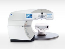 The same precise engineering has been used to develop the VisuMax femtosecond laser. The VisuMax is so precise that it requires 7 times less energy than other femtosecond lasers. Less energy is always key when treating the delicate tissue of the cornea. The high precision of the optics combined with the low energy create better outcomes for patients who are treated on the VisuMax femtosecond laser.
Gentle Treatment
Patient comfort was a high priority for the engineers at Carl Zeiss Meditec. Other femtosecond lasers use high suction during the creation of the corneal flap. This higher pressure can be uncomfortable and cause the patient to lose vision during the creation of the flap.
The VisuMax laser is an incredible advancement in patient comfort. The comfort provided by the VisuMax creates a much better surgical experience for the patient. The VisuMax laser used a curved surface like a contact lens. The curved surface is natural to the cornea and creates a comfortable experience for the patient. Patients that have had treatment on the VisuMax compare their experience to putting in a contact lens.
Doctors enjoy the increased safety when using the VisuMax for “bladeless” LASIK. Most potential complications can occur when using a mechanic blade to create the cornea flap. Doctors have embraced “bladeless” LASIK because of the increased patient safety and ease of use.
During treatment, the VisuMax femtosecond laser uses very precise optics to focus the laser energy. A femtosecond laser uses focused laser energy to create very small bubbles inside the cornea. The VisuMax places these tiny bubbles inside the cornea in a pattern that creates a corneal flap.
The doctor can create a very precise flaps that are unique to each patient. This flexibility and precision allow for a greater range of treatment options than are available with a mechanical blade.
Why do we Offer the VisuMax Laser for Bladeless All-Laser LASIK?
- Patient Safety
- Better outcomes
- Quicker recovery time
- Unsurpassed patient comfort
Flap creation with the VisuMax is a completely bladeless procedure. The creation of the corneal flap with the VisuMax is very fast and takes approximately 20 to 30 seconds, while the complete LASIK procedure may take approximately 15 minutes.
A very accurately focused laser beam is guided through to the cornea in a computerized treatment that is unique to each patient. The laser beam moves across and through the cornea, creating a layer of very tiny bubbles under its path. These bubbles quickly disappear, and the tissue above the bubbles becomes the corneal flap that can be easily lifted by your surgeon.
Everyone has a curved cornea. Nature often provides us with the best answers, and the VisuMax features a unique, curved contact glass to maximize patient comfort. This technology allows for the patient to maintain visual sight during the entire procedure. This is possible since the curved contact interface attaches to the cornea during the treatment and the cornea is only slightly flattened, which prevents unnecessarily high intraocular pressure and stress to the eye. This provides maximum comfort and it allows you to see throughout the entire procedure, unlike with microkeratomes or other femtosecond lasers, where your vision is “blacked out” for a while and patients have reported feeling uncomfortable pressure. Patients often describe the VisuMax procedure like the insertion of a soft contact lens.
Due to the Zeiss high performance optics, the laser beam is guided very precisely to the desired depth, resulting in most accurate flap thickness. In addition, the laser beam is directed in a very focused manner, which allows the doctor to apply only minimum laser energy to the eye during the treatment. As a result, the tissue outside the defined area of the cut remains untouched.
Since the VisuMax is able to create very thin corneal flaps, some patients who were previously not a candidate for LASIK due to thin corneas, are now benefiting from the procedure, thanks to the VisuMax. Your doctor will make the final determination.
Benefits of Blade-Free
The VisuMax laser provides more stability and greater flap precision when compared to LASIK. LASIK’s greatest complication is flap problems after surgery. Because blade free is so precise, this risk is significantly reduced. The blade-free Lasik vision correction surgery can produce real benefits for some patients.
They Include:
- Decreased risk of flap complications.
- Decreased incidence of postoperative dry eye
- Unlike other alternatives to LASIK, it preserves many of the benefits of LASIK including a short and largely pain-free recovery.
- More accurate outcomes in terms of patients’ postoperative vision.
- Benefits of vision correction surgery are available to those who might have previously been poor candidates.
- Less pressure on the eye during the surgery.
- Reduced incidence of patients needing a second operation to correct their vision.
- Flaps created with a laser are less likely to become dislodged later.
ZEISS MEL 80 EXCIMER LASER SYSTEM
ZEISS camera lenses were recognized as the world’s highest quality after their introduction in the 1800s. In the late 1980s, ZEISS optical lenses became the technology of choice for the developers of vision correction lasers.
Carl Zeiss became a notable manufacture of optics in Jena Germany in 1840. His company went on to develop revolutionary optics for microscopes and all types of optical devices. To this day, Zeiss is known worldwide as a leader in the manufacturing of highly precise quality optics. Zeiss utilized their extensive history in optics to develop their fourth generation Mel 80 excimer laser.
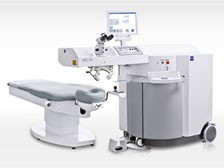 The MEL™ 80 is the latest excimer laser to receive FDA approval. The MEL™ 80 is a top quality Carl Zeiss Meditec product, designed to make the correction of vision defects, more patient-friendly and individual. All the parameters of this ultramodern work platform are oriented towards increasing efficiency, achieving optimum treatment results and the rapid recovery of vision. Key factors here are the very high speed treatments, and the high-performance eyetracker system that increases safety for patients.
- The fasted laser which leads to shorter treatment times and better outcomes.
- A lightning fast 1050 hertz eye tracker for a safer more accurate treatment.
- The latest laser cleared for FDA approval.
- A wavefront optimized treatment pattern that results in better correction and less glare at night time.
- A “flying spot” laser that utilizes a small treatment beam for a more accurate treatment profile and better vision.
Carl Zeiss Meditec has been a refractive surgery pioneer for more than 20 years. Combining its finest optics with the most advanced excimer laser technology available, Carl Zeiss Meditec brings you laser vision correction with a fourth-generation laser platform, the MEL 80.”MEL” stands for “Meditec Excimer Laser.”
The MEL 80 is a fourth generation Carl Zeiss excimer laser platform, taking vision correction to a new level. By reshaping the human cornea to its most natural shape during treatment, your vision can now be similar to the quality of lenses on which Carl Zeiss has built its reputation. The goal is to deliver high resolution vision with a custom treatment unique to your prescription. Carl Zeiss engineers created this advanced vision correcting technique using the latest wavefront techology innovations. This scientific method takes into consideration not only the clarity of vision from the shape of your eye, but it also treats other irregularities within your eye that may prevent you from having a higher quality of vision. One of the fastest excimer lasers in the world, your treatment is completed within seconds.
VISX STAR S4™ EXCIMER LASER SYSTEM
VISX laser systems have been used to perform millions of laser vision correction procedures in the U.S. and around the world. Every year, more doctors use VISX lasers to perform laser vision correction surgery than any other system. The VISX STAR S4 is one of the most advanced systems on the market today. This technologically advanced laser offers faster treatments using variable spot beam technology and an eye tracking system that tracks in all three dimensions.
The VISX STAR S4 can be used in combination with VISX CustomVue™ to address the unique imperfections of each individual’s vision. CustomVue can provide our patients with the potential to experience better vision than is possible with glasses or contact lenses.
Why We’ve Chosen the VISX STAR S4 Excimer Laser System:
- The STAR 4 variable beam technology allows the doctor to treat patients with different laser beam sizes and shapes, determined by each patient’s visual needs.
- The VISX Exclusive SmartBeam™ technology adjusts beam size according to treatment, minimizing the amount of corneal tissue that needs to be removed.
- With ActiveTrak™ 3D eye-tracking technology, the laser detects and compensates for small eye movements by guiding the laser beam to keep it centered precisely over the treatment area.
- VISX’s exclusive Active Trak™ is the only system that allows the doctor to track eye movement in all three dimensions. Having the beam centered correctly on the eye at all times helps ensure precise results.
- Unlike other lasers with eye-tracking technology, ActiveTrak requires no pupil dilation, leading to an even quicker recovery time.
- VISX laser systems produce an extremely smooth surface on the cornea after the procedure. Smoother ablations, as they’re known, promote faster healing and result in better visual outcomes.
- VISX laser systems achieve an optical zone that may result in reduced problems with night vision.
- VISX laser systems require a shorter procedure time than many lasers, which enhances comfort and may reduce the risk of postoperative complications.
BLADELESS INTRALASE™ FS LASER
The IntraLase FS Laser brings a new level of safety and assurance to vision correction surgery by providing an all-laser approach for optimal precision. Now, the accuracy of a computer-controlled laser may be used to create the corneal flap for LASIK, providing unprecedented control and the ability to customize the flap for each of our patients. Traditionally, the creation of a corneal flap was performed with a mechanical, hand-held device (known as a microkeratome) which moved across the cornea to cut the flap.
Because many LASIK complications are attributed to the unpredictable performance of the microkeratome, the new IntraLASIK all-laser surgical approach is being eagerly embraced across the US. Now, The IntraLase FS laser is a safer, computer-controlled alternative to a mechanical microkeratome for creating corneal flaps in the LASIK procedure. Although mechanical microkeratomes are safe, they can cause complications in 1 in 2500 or 5000 cases. Using the IntraLase FS Laser takes LASIK safety to the next level by significantly reducing the chance for removing that rare complication.
LASIK using the IntraLase FS Laser is called IntraLASIK. To make the corneal flap, the IntraLase FS Laser, the laser beam is focused into a tiny spot of energy that passes harmlessly through the outer layers of the cornea until reaching its exact focal point within the central layer of the cornea. There the corneal flap procedure is initiated, silently and painlessly, with remarkable precision. The IntraLase FS Laser can also be used to create the channels and entry cuts for placement of Intacs. Using the IntraLase FS Laser for this procedure assures that the two segments are placed at the exact same depth and removes the need for making the more surgically difficult mechanical channel.
Researchers are exploring the possibility of using the IntraLase FS Laser in other procedures, such as cornea transplants and glaucoma treatment.
Why We’ve Invested in the IntraLase FS Laser:
- IntraLASIK provides our patients with an extra margin of safety by eliminating potential complications from mechanical microkeratomes.
- IntraLASIK allows us to perform LASIK on patients with thin corneas because we can make a precisemore shallow flap, which allows us to correct higher levels of refractive error.
- IntraLASIK provides both the patient and the surgeon with a higher level of confidence knowing the creation of the flap will be without complications. Patients and surgeons relax more during the procedure making it more enjoyable for all.
- IntraLASIK makes laser vision correction an all laser procedure, which significantly adds to its precision and predictability.
- IntraLASIK also creates better fitting flaps. Once put back in place, these flaps form a tight seal, making flap dislocation after the procedure more difficult.
- The corneal flap procedure with IntraLASIK, using the IntraLase FS Laser is painless and more comfortable as there is much less pressure placed on the cornea with the laser vs the mechanical microkeratome.
Ziemer Galilei Topographer
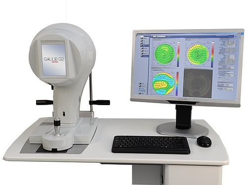
The GALILEI™ Dual Scheimpflug Analyzer is a high precision optical system for corneal topography and three-dimensional analysis of the anterior eye segment based on a revolving dual-channel Scheimpflug camera and a Placido Disk.
GALILEI™ combines the advantages of two technologies: Placido imaging furnishes high accuracy curvature data and Scheimpflug imaging is responsible for capturing precise elevation data. Precision Mapping of Cornea and Anterior Segment GALILEI™ incorporates several important diagnostic modalities. Combining them into a single device not only saves office space and investment cost – it also lays the basis for obtaining consistent and combined diagnostic information from a single set of merged measurement data.
GALILEI Integrated Functionalities:
- High-Resolution Scheimpflug images
- Pachymetry
- Corneal and Lens topography
- 3D Anterior chamber analysis
- Crystalline Lens thickness
- Corneal and lens densitometry
- Pupillometry
Bausch & Lomb Orbscan Topographer

ORBSCAN provides anterior segment data in the form of topographic surfaces. Elevation topography of the anterior cornea enables clinicians to more accurately visualize the shape of abnormal corneas, which leads to more accurate diagnoses and more consistent surgical results, at a much lower cost than most other topographers.
The reason for this is simple but important. When presented with maps of curvature or slope, you must mentally translate these data into elevation, because we visualize shape as topography (hills and valleys). This translation can be very difficult, often counter-intuitive if the curvature map is of an asymmetric or irregular surface. Curvature alone does not usually provide sufficient useable information for the mind to construct an accurate three-dimensional shape.
Traditionally, power maps (actually surface curvature) have been axis-centered, which are method dependent and only show one selected piece of the complete angle-dependent power. The new mean and toric decomposition of the complete curvature tensor, allow clinicians for the first time to see and quantify local curvature (and paraxial power) variations as well as the fundamental spherocylindrical components. Full corneal pachymetry allows surgeons to use regional blade settings, to measure the laser ablation depth from a fixed surface (the posterior cornea), and to assess the effect of the posterior surface on corneal power. Accurate knowledge of both surfaces will provide researchers with real data for corneal mechanics simulations. The point of minimum corneal thickness is a local center of axisymmetry, which for normal corneas aligns closely to the overall optical axis of the eye.
Corneal Topography
Corneal topography is a computer-assisted diagnostic tool that creates a three-dimensional map of the surface curvature of the cornea. The cornea (the front window of the eye) is responsible for about 70 percent of the eye’s focusing power. An eye with normal vision has an evenly rounded cornea, but if the cornea is too flat, too steep, or unevenly curved, less than perfect vision results. The greatest advantage of corneal topography is its ability to detect irregular conditions invisible to most conventional testing.
Corneal topography produces a detailed, visual description of the shape and power of the cornea. This type of analysis provides your doctor with very fine details regarding the condition of the corneal surface. These details are used to diagnose, monitor, and treat various eye conditions. They are also used in fitting contact lenses and for planning surgery, including laser vision correction. For laser vision correction the corneal topography map is used in conjunction with other tests to determine exactly how much corneal tissue will be removed to correct vision and with what ablation pattern.
Computerized corneal topography can be beneficial in the evaluation of certain diseases and injuries of the cornea including:
- Corneal diseases
- Corneal abrasions
- Corneal deformities
- Irregular astigmatism following corneal transplants
- Postoperative cataract extraction with acquired astigmatism
The corneal topography equipment consists of a computer linked to a lighted bowl that contains a pattern of rings. During a diagnostic test, the patient sits in front of the bowl with his or her head pressed against a bar while a series of data points are generated. Computer software digitizes these data points to produce a printout of the corneal shape, using different colors to identify different elevations, much like a topographic map of the earth displays changes in the land surface. The non-contact testing is painless and brief.